Case ID: 1186
Publication date: 05 Feb, 2016
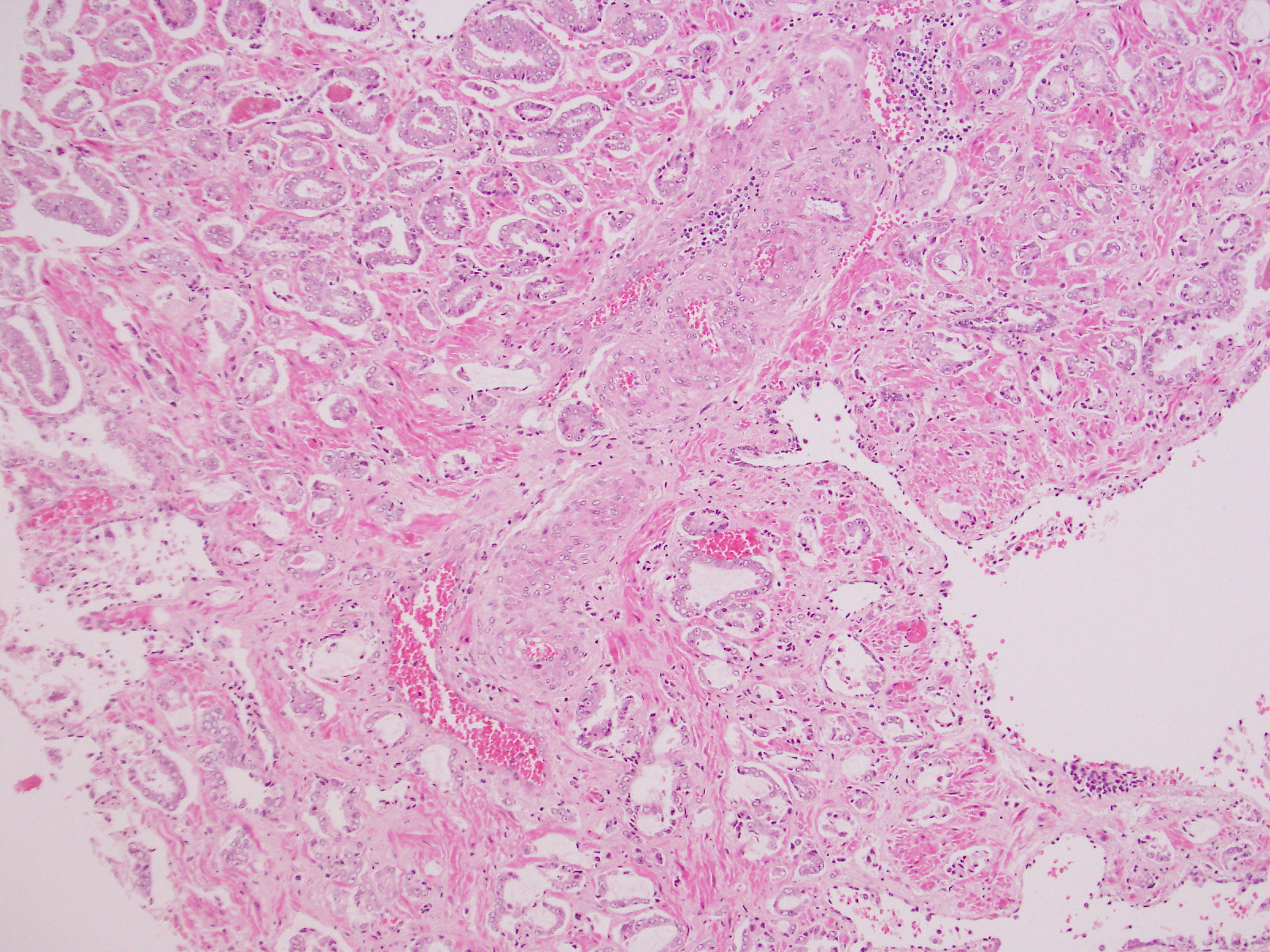
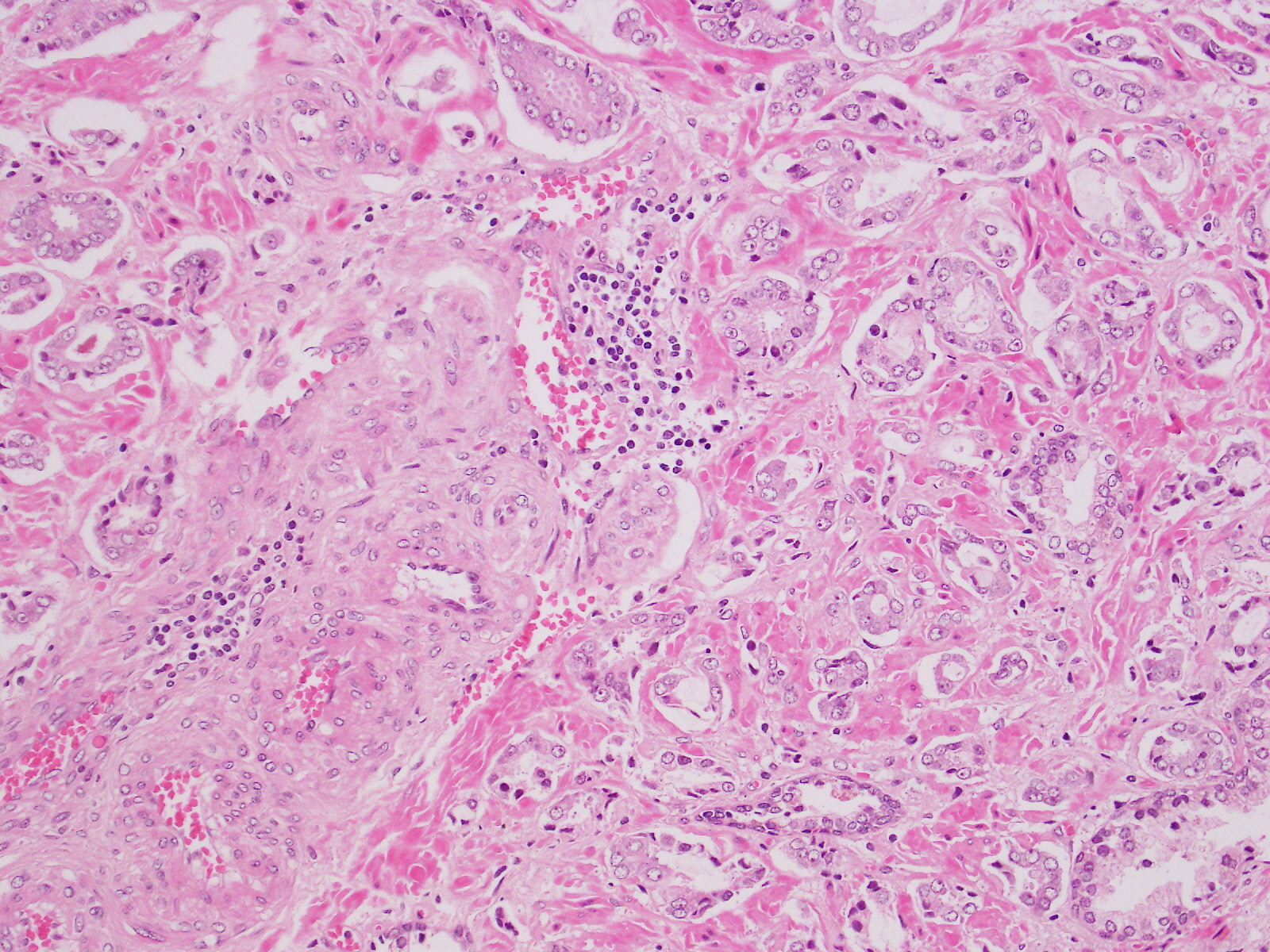
Consensus grade: GS 3+3=6 (ISUP 1)
Show diagnosis by expert panel members| User | Diagnosis | Difficulty | Comment |
|---|---|---|---|
| Pathologist 1 | GS 3+3=6 (ISUP 1) | Typical | |
| Pathologist 2 | GS 3+3=6 (ISUP 1) | Typical | |
| Pathologist 3 | GS 3+3=6 (ISUP 1) | Typical | |
| Pathologist 4 | GS 3+4=7 (ISUP 2) | Typical | |
| Pathologist 5 | GS 3+3=6 (ISUP 1) | Borderline higher |
Focal tangential sectioning of small gland pattern 3, insufficient for cluster of poorly formed glands of pattern 4. |
| Pathologist 6 | GS 3+3=6 (ISUP 1) | Borderline lower |
Have seen this case before |
| Pathologist 7 | GS 3+3=6 (ISUP 1) | Typical | |
| Pathologist 8 | GS 3+3=6 (ISUP 1) | Typical | |
| Pathologist 9 | GS 3+3=6 (ISUP 1) | Borderline higher | |
| Pathologist 10 | Other | Typical |
3+5=8 |
| Pathologist 11 | GS 3+3=6 (ISUP 1) | Borderline higher | |
| Pathologist 12 | GS 3+4=7 (ISUP 2) | Typical | |
| Pathologist 13 | GS 3+3=6 (ISUP 1) | Typical | |
| Pathologist 14 | GS 3+4=7 (ISUP 2) | Borderline higher |
Again, retraction artifact |
| Pathologist 15 | GS 3+4=7 (ISUP 2) | Borderline lower | |
| Pathologist 16 | GS 3+3=6 (ISUP 1) | Typical |
This image was also used as the high power image in Case ID 1210 GK57 |
| Pathologist 17 | GS 3+3=6 (ISUP 1) | Borderline higher | |
| Pathologist 18 | GS 3+3=6 (ISUP 1) | Borderline higher | |
| Pathologist 19 | GS 3+4=7 (ISUP 2) | Typical | |
| Pathologist 20 | GS 3+3=6 (ISUP 1) | Typical |
This is an exact repeat of a case that's numbered in the 60's or 70's. |
| Pathologist 21 | GS 3+4=7 (ISUP 2) | Borderline lower | |
| Pathologist 22 | GS 3+3=6 (ISUP 1) | Typical | |
| Pathologist 23 | GS 3+3=6 (ISUP 1) | Typical |
but considering the nucleoli 3 couild be an option |
Case description (by case creator):