Case ID: 774
Publication date: 28 Aug, 2015
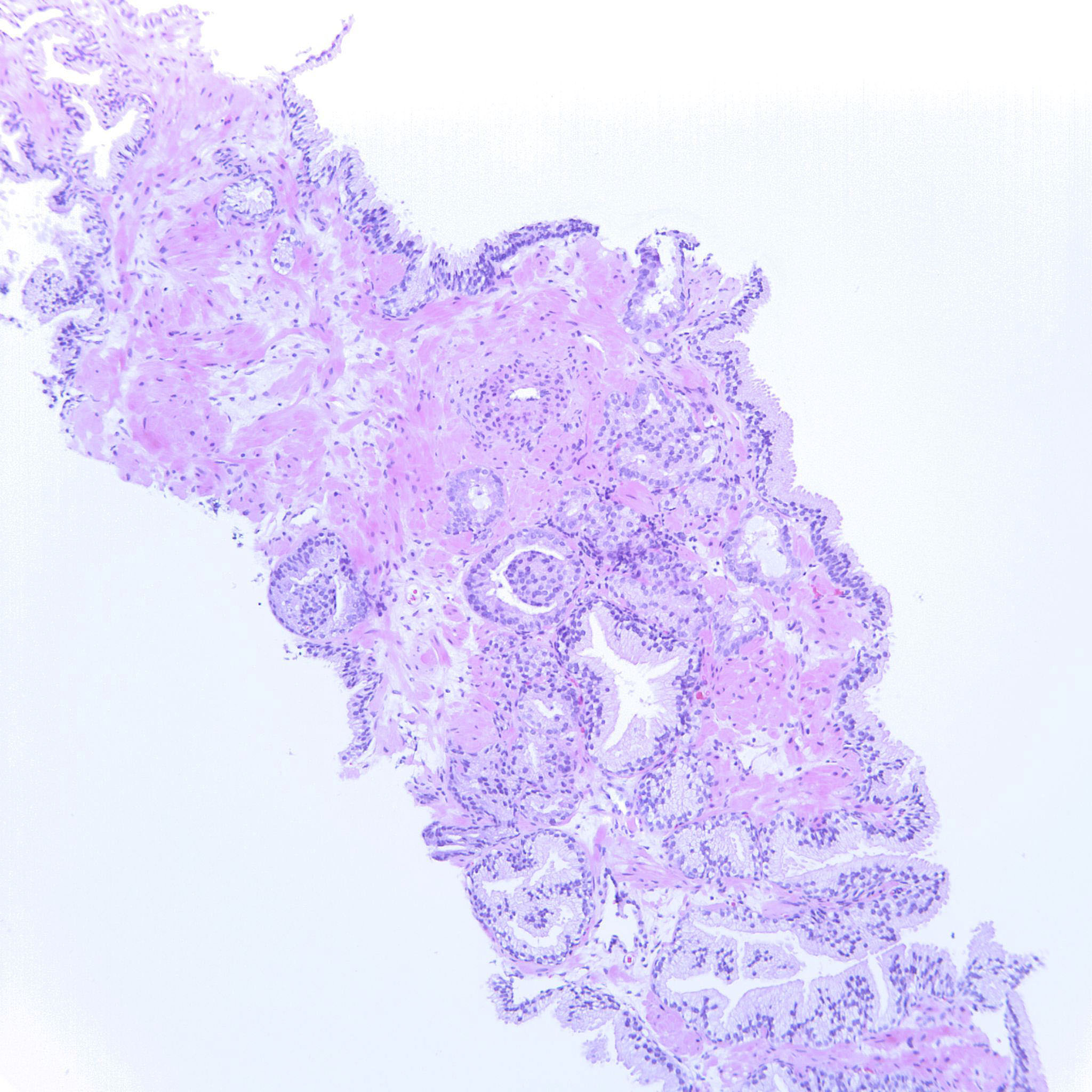
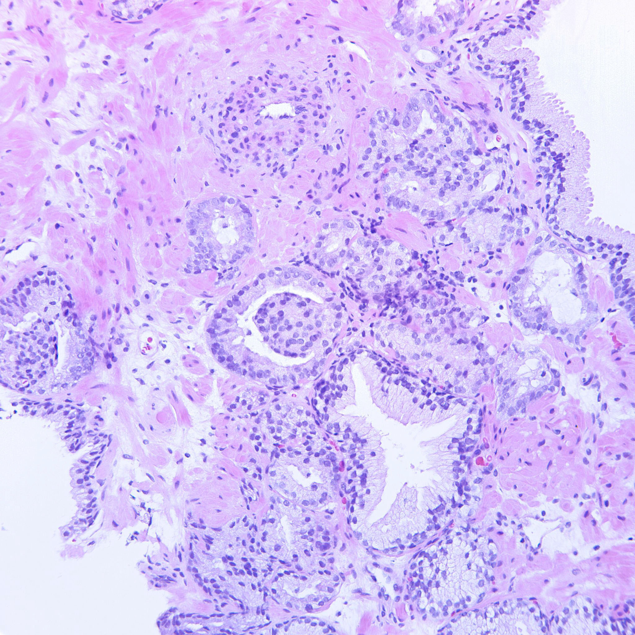
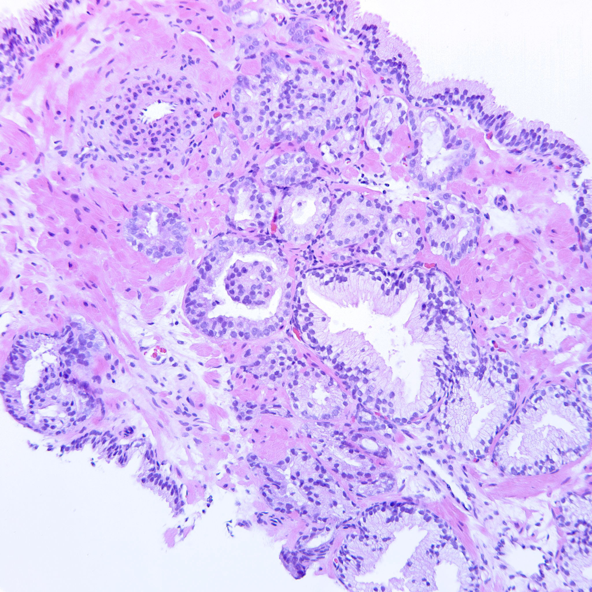
Consensus grade: GS 3+4=7 (ISUP 2)
Show diagnosis by expert panel members| User | Diagnosis | Difficulty | Comment |
|---|---|---|---|
| Pathologist 1 | GS 3+4=7 (ISUP 2) | Borderline higher | |
| Pathologist 2 | GS 3+3=6 (ISUP 1) | Typical |
Glomeruloid is 3+3.First description was grade 3, NOT grade 4-- WHO 2015 is wrong, but the dominant voice at the meeting always gets his way, right? |
| Pathologist 3 | GS 4+3=7 (ISUP 3) | Borderline lower | |
| Pathologist 4 | GS 3+4=7 (ISUP 2) | Borderline lower | |
| Pathologist 5 | GS 3+4=7 (ISUP 2) | Typical | |
| Pathologist 6 | GS 3+4=7 (ISUP 2) | Typical | |
| Pathologist 7 | GS 3+3=6 (ISUP 1) | Borderline higher |
mostly discrete glands, one glomeruloid feature, still compatible with G3 |
| Pathologist 8 | GS 3+4=7 (ISUP 2) | Typical | |
| Pathologist 9 | GS 3+3=6 (ISUP 1) | Borderline higher | |
| Pathologist 10 | GS 3+4=7 (ISUP 2) | Typical | |
| Pathologist 11 | GS 4+3=7 (ISUP 3) | Borderline higher | |
| Pathologist 12 | GS 3+4=7 (ISUP 2) | Typical | |
| Pathologist 13 | GS 4+3=7 (ISUP 3) | Typical |
75% pattern 4 |
| Pathologist 14 | GS 3+4=7 (ISUP 2) | Typical | |
| Pathologist 15 | GS 4+3=7 (ISUP 3) | Borderline lower | |
| Pathologist 16 | GS 3+4=7 (ISUP 2) | Typical | |
| Pathologist 17 | GS 3+4=7 (ISUP 2) | Borderline lower | |
| Pathologist 18 | GS 3+4=7 (ISUP 2) | Typical |
The 2nd HP image is not represented in the 1st LP image? |
| Pathologist 19 | GS 3+4=7 (ISUP 2) | Typical | |
| Pathologist 20 | GS 3+4=7 (ISUP 2) | Typical | |
| Pathologist 21 | GS 3+4=7 (ISUP 2) | Borderline higher |
Am reluctant to upgrade on such limited foci. Are the last 2 pics same area in different levels? |
| Pathologist 22 | GS 3+4=7 (ISUP 2) | Typical |
annoying ase, does not look too bad, but lots of glomeruloid aspects... |
| Pathologist 23 | GS 4+3=7 (ISUP 3) | Typical | |
| Pathologist 24 | GS 3+4=7 (ISUP 2) | Typical |
Case description (by case creator):
Most of the cancer is GP3 but there are also some glomeruloid bodies justifying GP4. The two 20x fields are from two adjacent sections of the same biopsy, in order to verify that it is not a tangential cut.