Case ID: 149
Publication date: 05 Oct, 2015
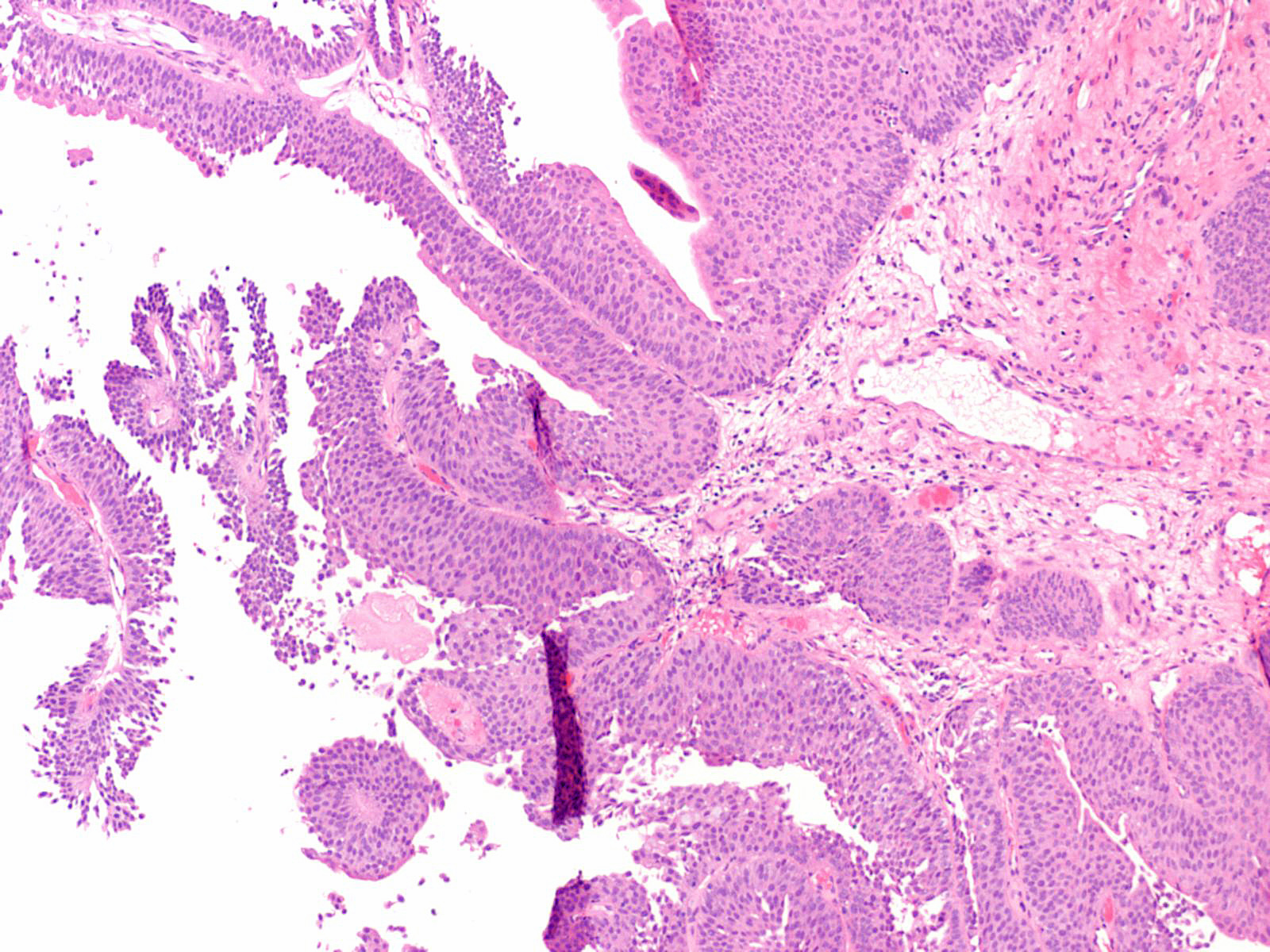
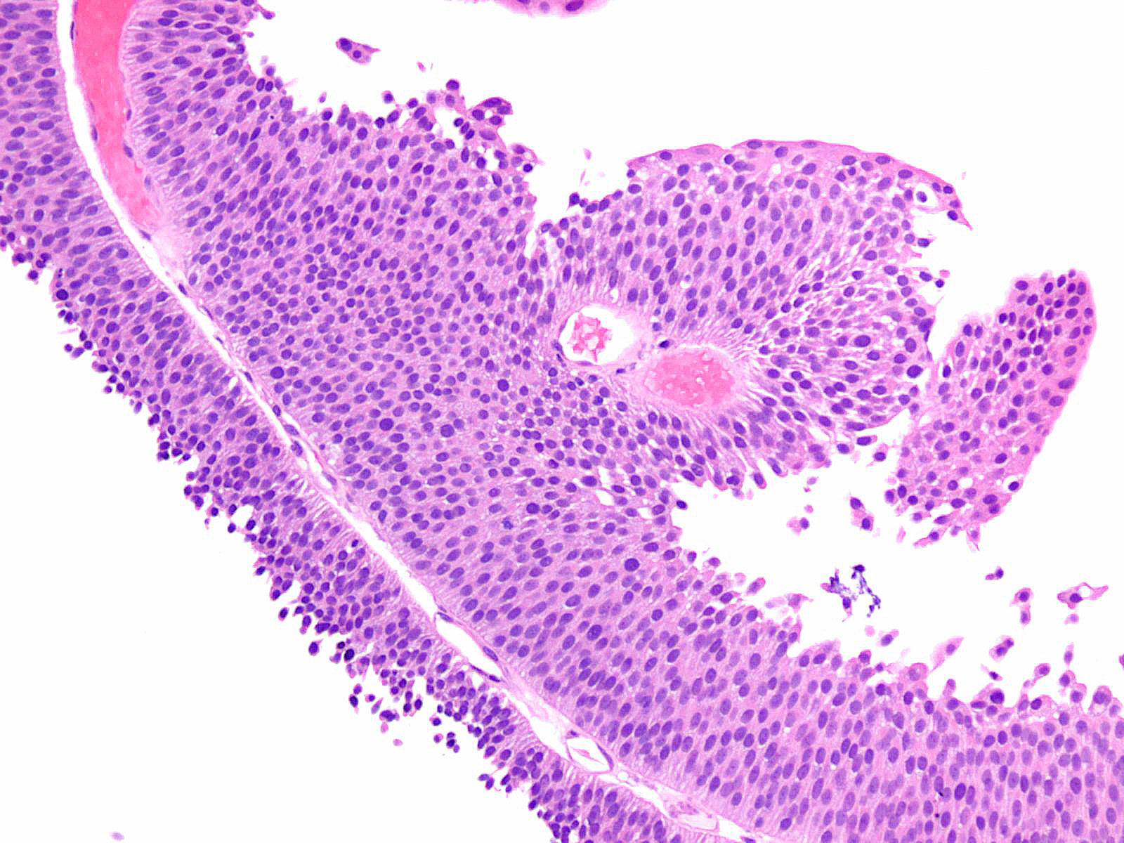
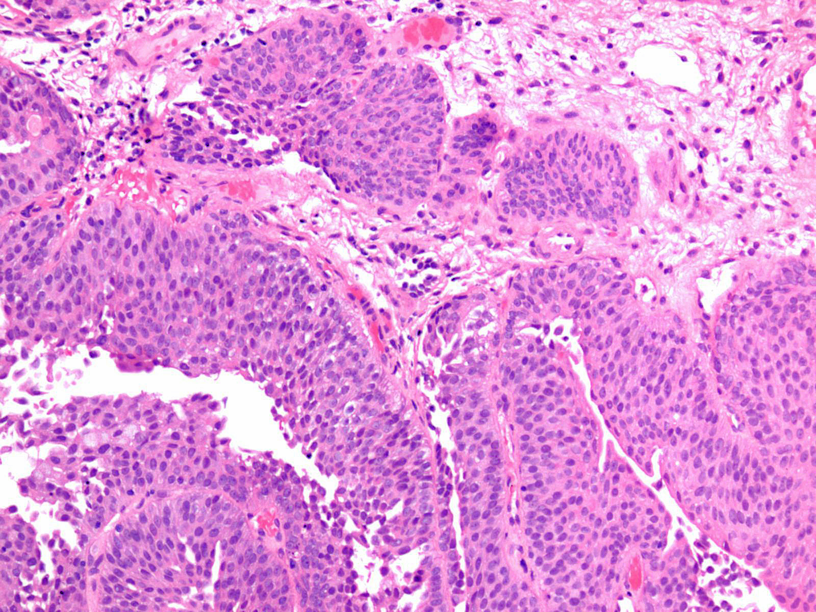
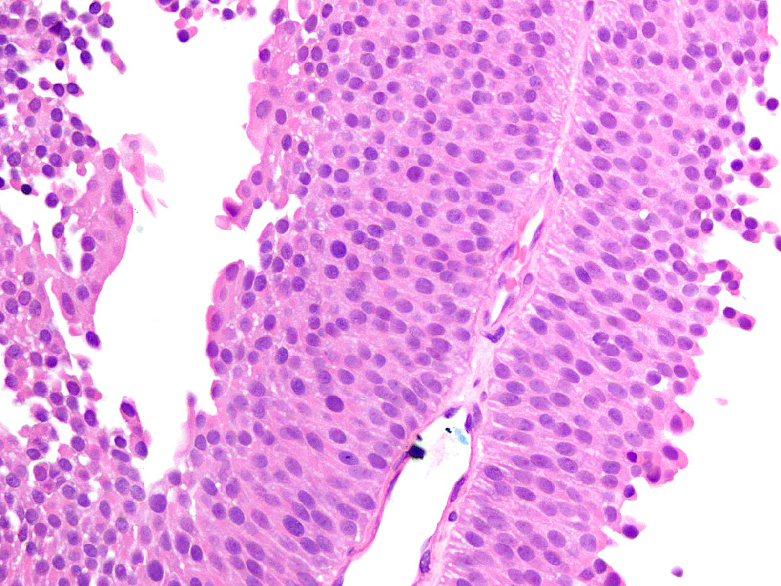
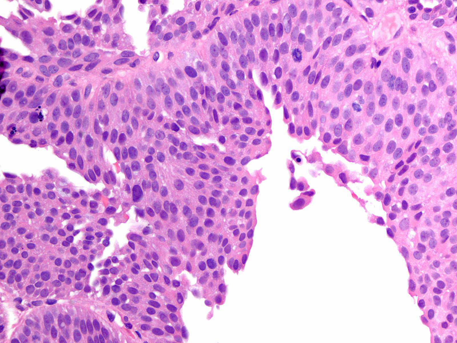
Consensus grade: Low-grade papillary urothelial carcinoma (LG-PUC)
Show diagnosis by expert panel members| User | Diagnosis | Difficulty | Comment |
|---|---|---|---|
| Pathologist 1 | Low-grade papillary urothelial carcinoma (LG-PUC) | Typical | |
| Pathologist 2 | High-grade papillary urothelial carcinoma (HG-PUC) | Typical | |
| Pathologist 3 | Low-grade papillary urothelial carcinoma (LG-PUC) | Borderline higher | |
| Pathologist 4 | Low-grade papillary urothelial carcinoma (LG-PUC) | Typical | |
| Pathologist 5 | High-grade papillary urothelial carcinoma (HG-PUC) | Typical | |
| Pathologist 6 | Low-grade papillary urothelial carcinoma (LG-PUC) | Typical | |
| Pathologist 7 | Low-grade papillary urothelial carcinoma (LG-PUC) | Typical | |
| Pathologist 8 | Low-grade papillary urothelial carcinoma (LG-PUC) | Typical | |
| Pathologist 9 | Low-grade papillary urothelial carcinoma (LG-PUC) | Typical | |
| Pathologist 10 | Low-grade papillary urothelial carcinoma (LG-PUC) | Typical | |
| Pathologist 11 | Low-grade papillary urothelial carcinoma (LG-PUC) | Typical |
No specific comment. |
| Pathologist 12 | Low-grade papillary urothelial carcinoma (LG-PUC) | Typical | |
| Pathologist 13 | Low-grade papillary urothelial carcinoma (LG-PUC) | Typical | |
| Pathologist 14 | High-grade papillary urothelial carcinoma (HG-PUC) | Bordering on lower | |
| Pathologist 15 | PUNLMP | Typical | |
| Pathologist 16 | Low-grade papillary urothelial carcinoma (LG-PUC) | Bordering on higher | |
| Pathologist 17 | Low-grade papillary urothelial carcinoma (LG-PUC) | Typical | |
| Pathologist 18 | Low-grade papillary urothelial carcinoma (LG-PUC) | Typical | |
| Pathologist 19 | Low-grade papillary urothelial carcinoma (LG-PUC) | Typical | |
| Pathologist 20 | PUNLMP | Typical | |
| Pathologist 21 | Low-grade papillary urothelial carcinoma (LG-PUC) | Typical | |
| Pathologist 22 | Low-grade papillary urothelial carcinoma (LG-PUC) | Bordering on higher |
Mostly still LG. |
Case description (by case creator):
Lesion shows mild to moderate variation in nuclear size, shape and chromatin. Scattered nuclei are significantly enlarged and hyperchromatic relative to other nuclei. Lesion maintains an overall orderly appearance. Few mitoses are noted.