Case ID: 550
Publication date: 11 Jan, 2016
Consensus grade: Other
Show diagnosis by expert panel members| User | Diagnosis | Difficulty | Comment |
|---|---|---|---|
| Pathologist 1 | Other | Typical | |
| Pathologist 2 | Other | Typical |
AML with cysts |
| Pathologist 3 | Other | Typical |
AMELC |
| Pathologist 4 | Other | Typical | |
| Pathologist 5 | Other | Typical | |
| Pathologist 6 | Other | Not typical |
I need immunohistochemistry to diagnose this case |
| Pathologist 7 | Other | Not typical | |
| Pathologist 8 | Other | Not typical | |
| Pathologist 9 | Other | Typical | |
| Pathologist 10 | Other | Typical | |
| Pathologist 11 | Other | Typical |
AML |
| Pathologist 12 | Other | Typical | |
| Pathologist 13 | Other | Typical | |
| Pathologist 14 | Other | Typical |
AML |
| Pathologist 15 | Insufficient tumor for diagnosis | Typical |
angiomyolipoma with epithelial cyst (dr Fine´s variant) |
| Pathologist 16 | Other | Typical |
AMLEC |
| Pathologist 17 | Other | Not typical |
AML with epithelial cyst (AMLEC)... good case |
| Pathologist 18 | Other | Typical |
AML |
| Pathologist 19 | Other | Typical |
AML with epithelial cyst |
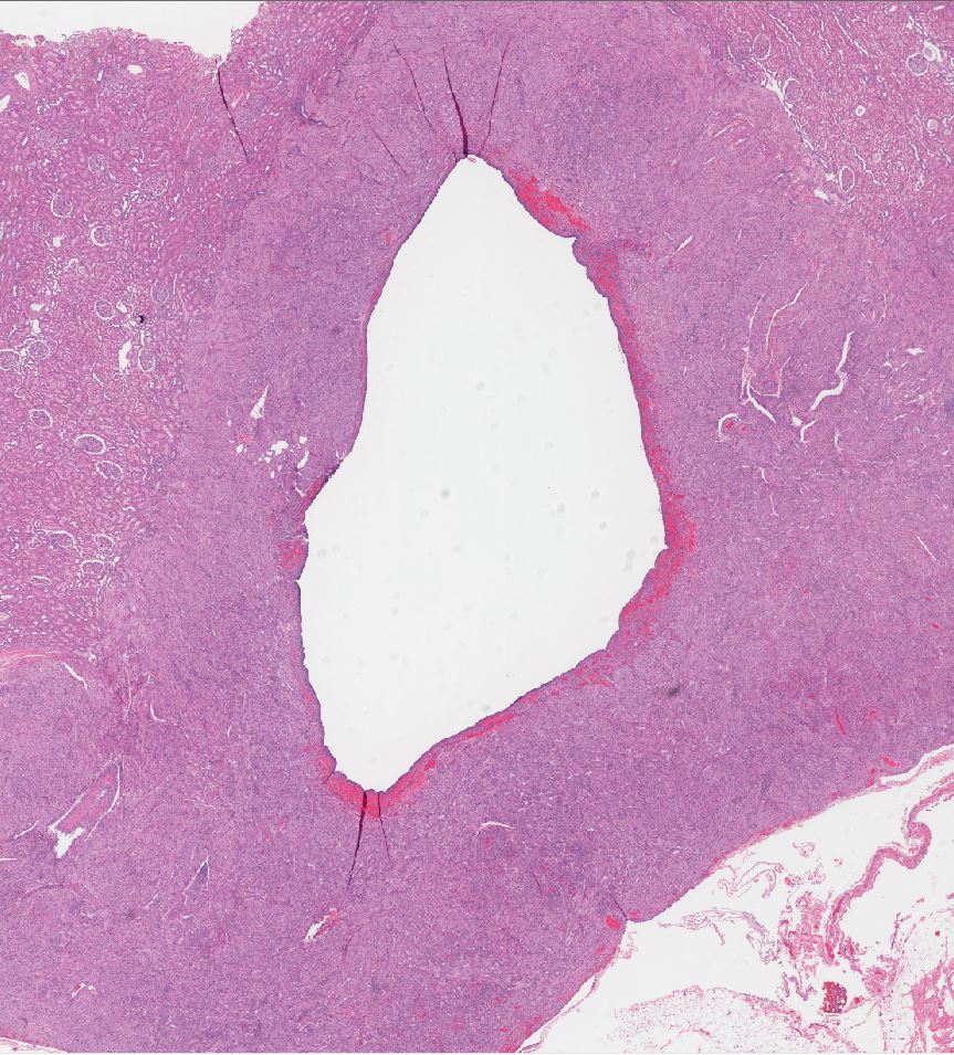
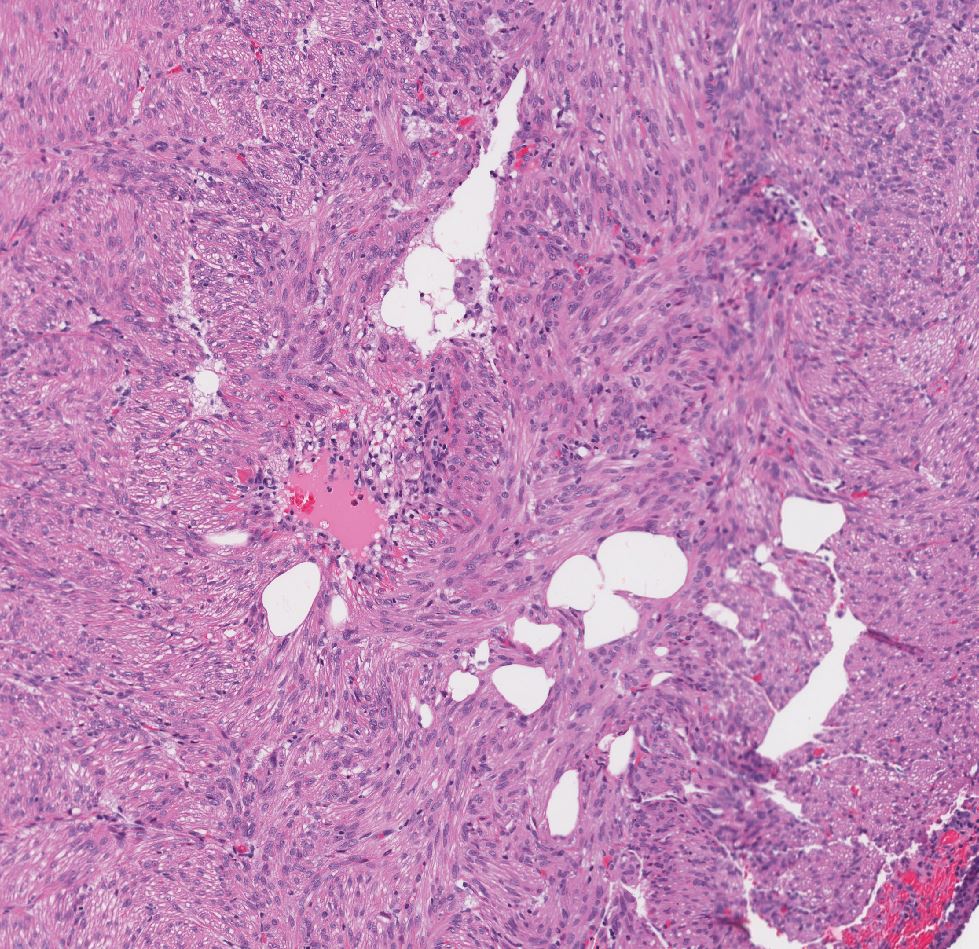
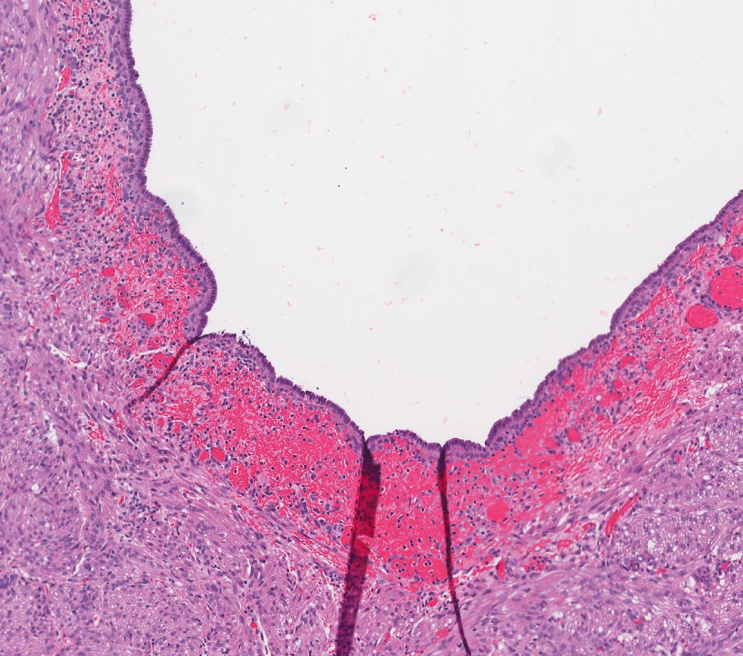
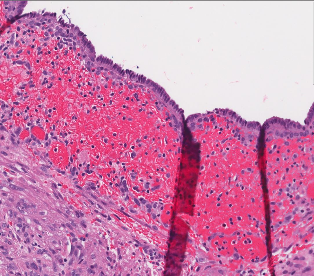
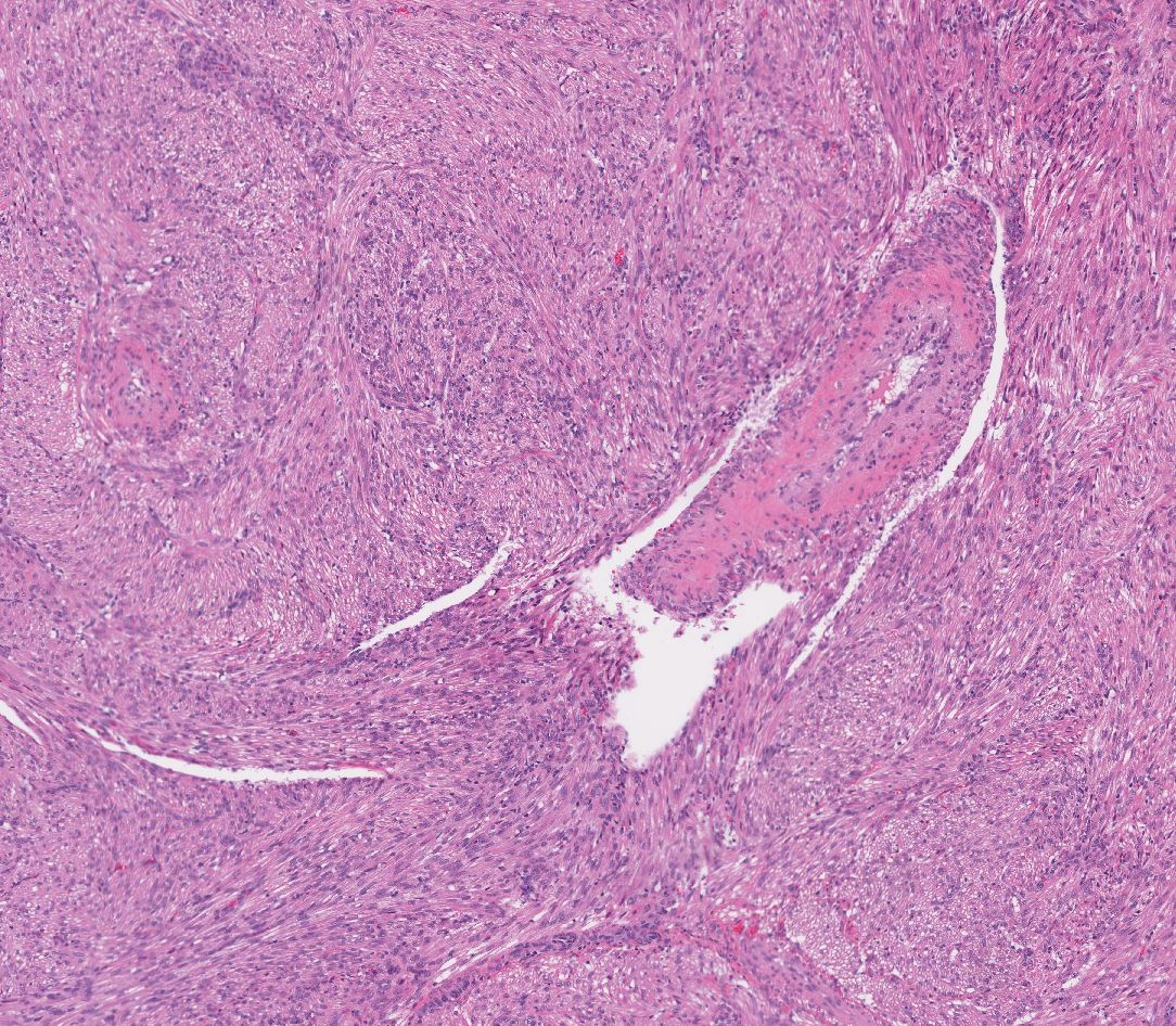
Case description (by case creator):
Renal angiomyolipoma with epithelial cyst (AMLEC). The spindle cells surrounding the central cyst are positive for HMB45 and Melan A. The lesion also contains scattered, thick-walled blood vessels and microscopic areas of admixed fat. The central cyst is lined by a single layer of bland cuboidal/hobnail epithelial cells.