Case ID: 1127
Publication date: 25 Jan, 2016
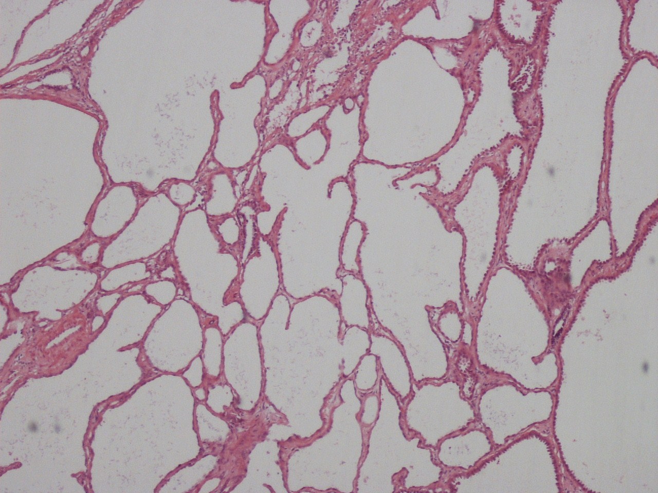
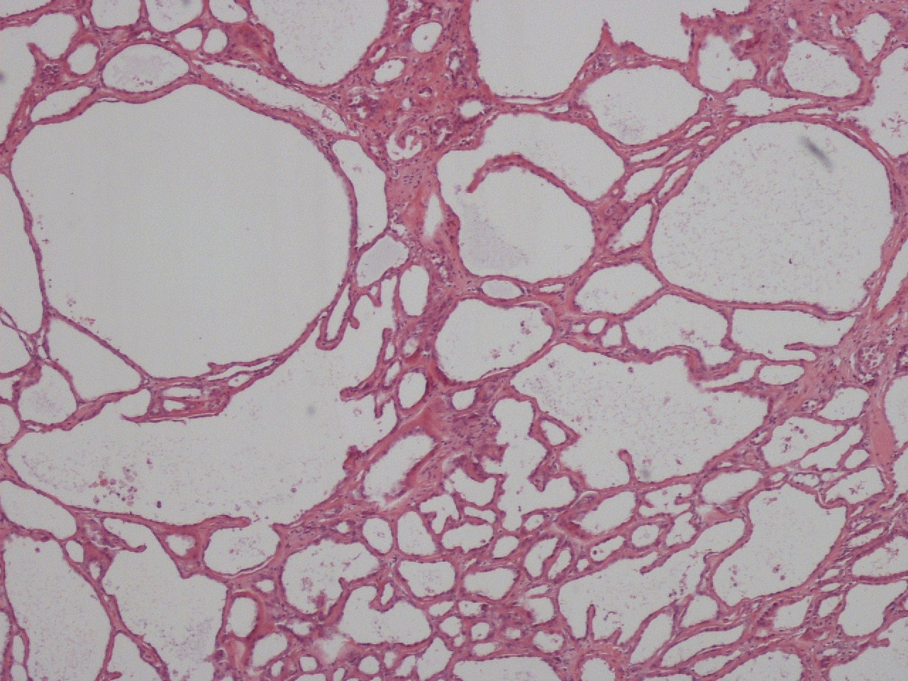
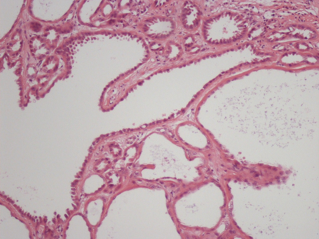
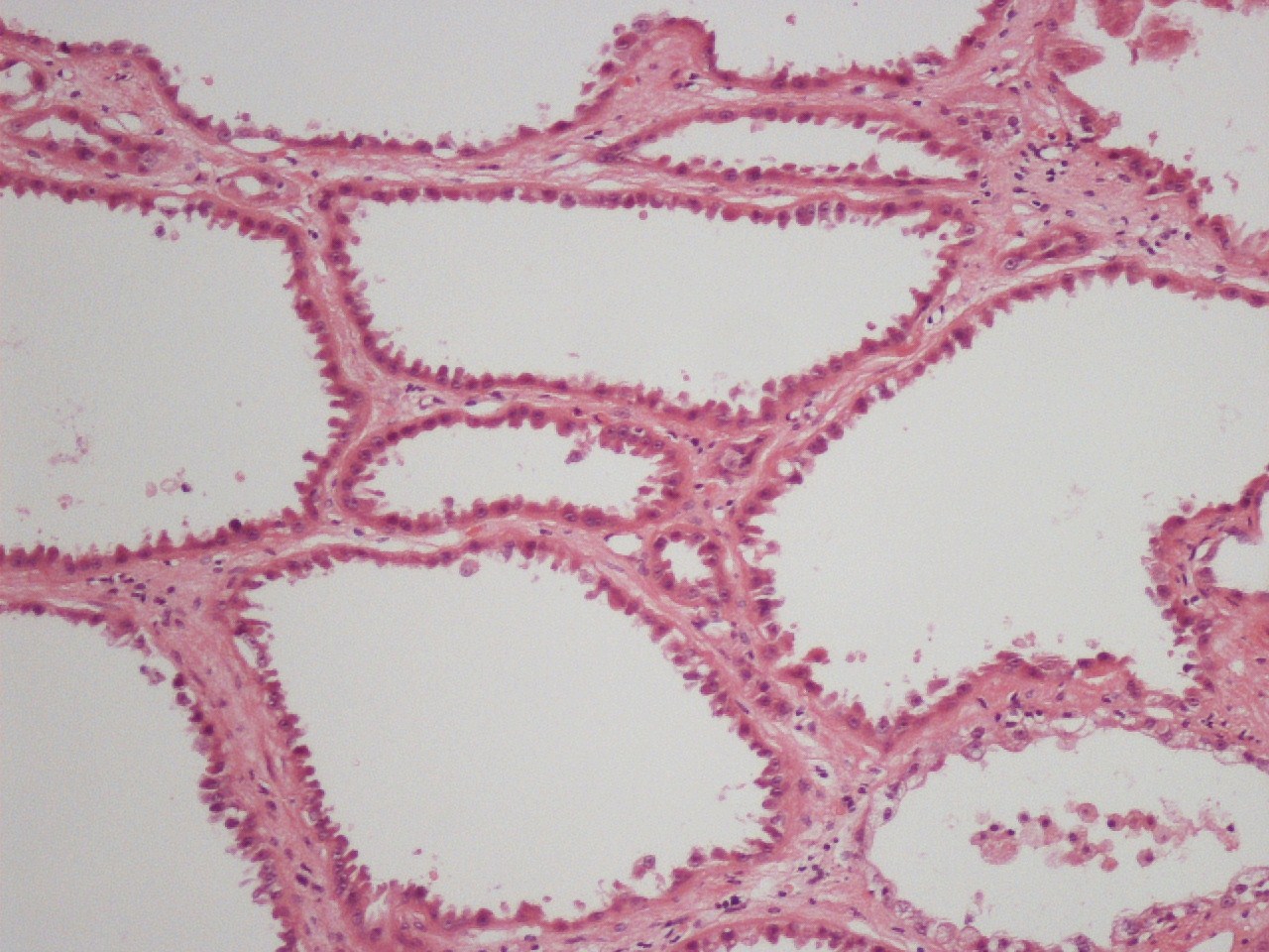
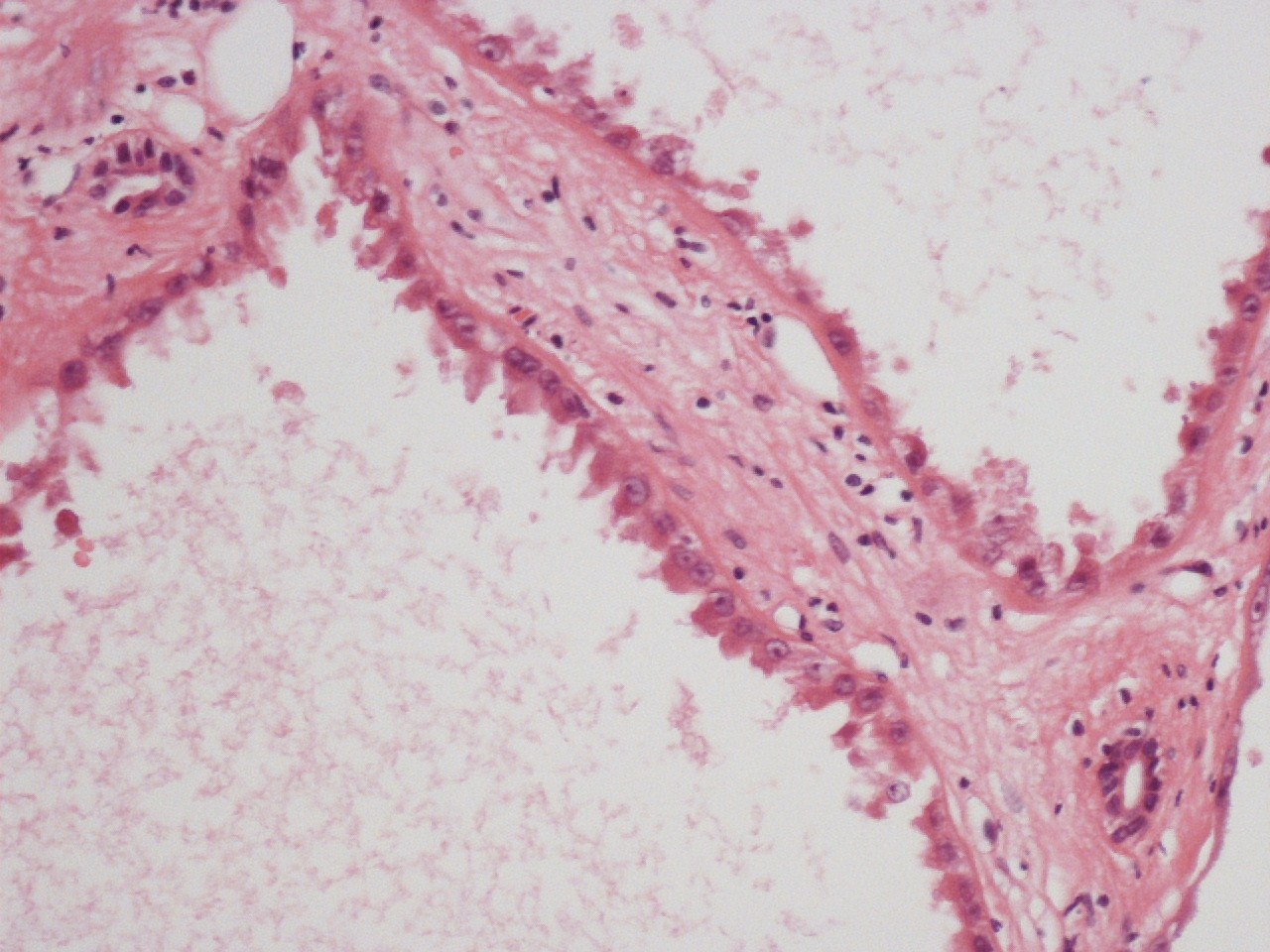
Consensus grade: Tubulocystic RCC
Show diagnosis by expert panel members| User | Diagnosis | Difficulty | Comment |
|---|---|---|---|
| Pathologist 1 | Tubulocystic RCC | Typical | |
| Pathologist 2 | Tubulocystic RCC | Typical |
HLRCC needs to ruled out by ancillary studies before the above diagnosis is rendered. |
| Pathologist 3 | Tubulocystic RCC | Typical | |
| Pathologist 4 | Tubulocystic RCC | Typical |
Borderline quality - not a great H&E |
| Pathologist 5 | Tubulocystic RCC | Typical | |
| Pathologist 6 | Tubulocystic RCC | Typical | |
| Pathologist 7 | Tubulocystic RCC | Typical | |
| Pathologist 8 | Tubulocystic RCC | Typical | |
| Pathologist 9 | Tubulocystic RCC | Typical | |
| Pathologist 10 | Tubulocystic RCC | Typical | |
| Pathologist 11 | Tubulocystic RCC | Typical | |
| Pathologist 12 | Tubulocystic RCC | Typical | |
| Pathologist 13 | Tubulocystic RCC | Typical | |
| Pathologist 14 | Tubulocystic RCC | Typical | |
| Pathologist 15 | Tubulocystic RCC | Typical | |
| Pathologist 16 | Tubulocystic RCC | Typical | |
| Pathologist 17 | Tubulocystic RCC | Typical |
Case description (by case creator):
Renal tumor from a 48 year old male. The tumor was 7.3cms in diameter and had a grey spongy morphology. It is composed of cysts of varying sizes. These are lined by a single layer of tumor cells. these are variously cuboidal, columnar or on occasion hob-nailed. The cytoplasm is eosinophilic and faintly granular. Nuclei are centrally placed and although showing minimal pleomorphism do have prominent single nucleoli. Mitoses are rare.